Carbohydrates, lipids, proteins, and nucleic acids are known as four biological macromolecules, which are closely related to fertilization, development, immune and nervous system formation, cell canceration and other life phenomena. Intracellular carbohydrates are involved in a wide range of physiological activities. The prediction and identification of glycan structures are very important in the diagnosis and pathology of diseases in carbohydrate metabolism. The identification of the structure of the sugar chain requires professional, complex, mature and stable technology compared with protein and nucleic acid.
For a long time, the research on glycan structure prediction has focused on the N-glycostructure of host cells expressed by recombinant proteins, such as CHO based on kinetic and statistical models. Gene diagnosis technology has been one of the most popular detective technologies in the field of life science recently. With the development of high-throughput sequencing technology, it is possible to use RNA-seq data for genetic diagnosis and disease screening. Therefore, it is quite necessary to develop a more convenient tool to predict the structure of glycans in the research for the detection of human diseases and the mutual regulation of glycans in cells.
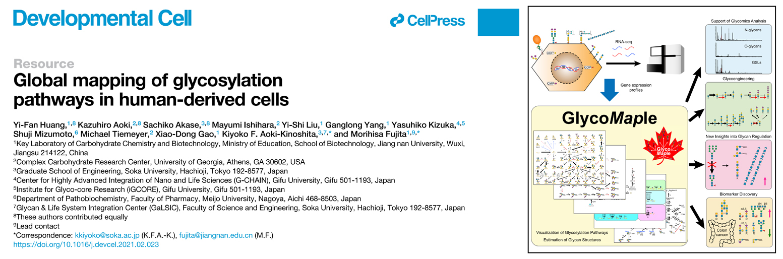
On March 16, 2021, Global mapping of glycosylation pathways in human-derived cells was published in the Developmental Cell by Huang Yi-fan in Prof.Morihisa Fujita group, School of Bioengineering, Jiangnan University, in collaboration with Kiyoko Aoki-Kinoshita, Soka University, Japan, and Kazuhiro Aoki, University of Georgia, USA. Glycomaple, a predictive tool based on RNA-seq data to visualize the glycan metabolic pathway and evaluate the synthesis potential of glycans to predict the glycan structures. In the N-glycan deficient cell library of the HEK293 cell line, we used a predictive tool combined with a multi-omics study to reveal the global glycan structural changes induced by the transcriptional regulation of multiple glycosylation pathways in the HEK293 cells with dominant synthetic high mannose type N-glycans (Figure 1).
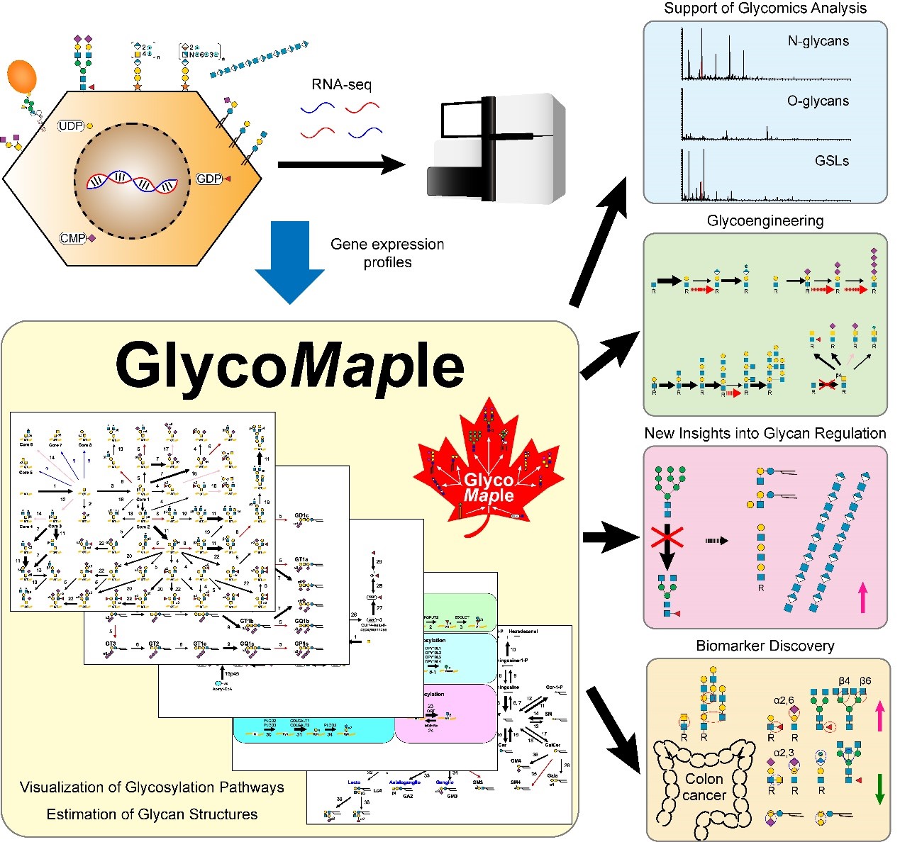
Figure 1. Overview of Glycomaple
The author firstly based on the literature collected 950 glycosylation related genes, a comprehensive list of contains CAZy database registration code 200 glycosyltransferase, 78 glycoside hydrolase and 15 sugar-binding protein of human genes, and coding sulfotransferases, regulating factor, the synthesis and sugar nucleotide synthesis and transport, and genes involved in Golgi homeostasis for glycoconjugate synthesis and degradation. Based on the gene encoding protein catalytic activity, 19 glycosylation silico pathways were drawn to visualize metabolic processes.
To investigate whether RNA-seq data can be directly applied to the prediction of glycan structures, the authors first performed a statistical assessment based on 53 possible glycan structures of the O-glycosynthesis pathway. The AUC of model cell HEK293 under the ROC curve was 0.90, demonstrating the feasibility of using transcriptional information for prediction (Figure 2).
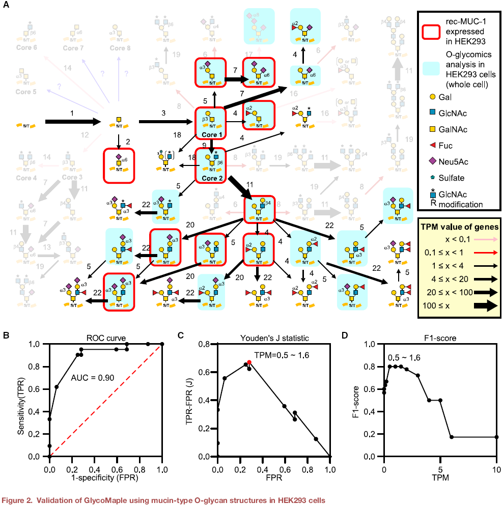
Figure 2. Evaluation of GlycoMaple using mucin-type O-sugar structure in HEK293 cells
Then, the glycosylation capacity of HEK293 cells was evaluated by glycosomic based analysis of the N-glycans, O-glycans and glycolipids of HEK293 cells. By integrating the cDNA of glycosyltransferase, the wild-type HEK293 cells were given the ability to express terminal modifications such as HNK-1, poly-sialic acid and I-type branched polylactosamine. On the other hand, the expression of the LacdiNAc structure was eliminated by knockout of key genes with CRISPR/Cas9. It was found that the genes with the lowest transcriptional level on the glycan biosynthesis pathway restricted the expression of the final glycan structures.
The author used the CRISPR/Cas9 to construct a cell bank containing 40 N-glyco-deficient HEK293 cell lines. In lectin-based glycosomics studies, a significant cluster of cell lines that mainly expressed high-mannose N-glycans were observed. An abnormal accumulation of Lactosamine modifications on the surface of these clustered cell lines appeared beyond expectation. Since the Lactosamine motif was primarily modified on the terminal of N-glycans, while no associated modification sites were possible on high-mannose N-glycans. In this case, seven representative knockout cell lines were subsequently subjected to multi-omics studies. It was found that the composition of the glycan structures on glycolipids of the cell lines expressing high mannose type N-glycans was significantly altered, with a significant increase in the Lactosamine modification, and poly-lactosamine modified glycolipids. B3GnT5, a key gene which encodes LC3 glycolipid synthetase in the glycolipid pathway, was knockout in the mannotransferase I-deficient cell line, T-KO cells, therewith the Lactosamine modification on the surface disappeared, demonstrating that the accumulated Lactosamine modification occurs on glycolipids. Transcriptome analysis in GlycoMaple revealed significant upregulation of several glyco-related genes such as CERS1, HAS2 and RENBP in T-KO cells. Whereafter lipid composition and the sugar nucleotides substrate were analyzed, a new regulatory mechanism was found. In the T-KO cells which mainly express high mannose type N-glycans, UDP-GlcNAc substrate was accumulated. Cells active an adaptive transcription regulation by the increase of lipid biosynthesis, the promotion of the UDP-GlcNAc utilization in the lipid part, and the evaluated production of hyaluronic acid to extracellular space, in order to release the substrate stress caused by N-glycan deficiency. Previous studies have found that in a variety of cancerous cells, the accumulation of high-mannose N-glycans and changes in lipid components, as well as the upregulation of hyaluronic acid synthesis, were observed due to cellular metabolic stress. This study provides a potential explanation for the transcriptional regulation of this phenomenon.
In the end, the author develops a network tool, which can be widely used in the cell research of various human tissues and organs. RNA-Seq data for 64 human-derived cell lines and 37 human tissue types were integrated into the web tool. In the cluster analysis of human tissue transcriptome, the transcriptome pattern of digestive tract organs tended to be similar, revealing that the high expression of key proteins in the synthesis of O-glycans in digestive organs, contributed to the richness and complexity of O-glycans. Besides, the transcriptome sequencing data of 288 diseased tissue samples from colon cancer patients in the TCGA database and 304 healthy tissue samples in the GTEX database were compared and analyzed using the comparison function of the network tool. The changes in the glycan structures during the process of colon cancer degeneration were successfully predicted (Figure 3). This work fills a gap in the field of glycobiology in predicting the structure of glycans by using gene transcriptional information.
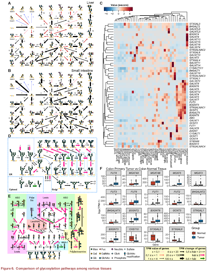
Figure 3. Comparison of glycosylated metabolic pathways in different tissue types
In summary, this research used RNA-seq data to visualize glycan metabolic pathways and verified the reliability of using the transcription data to predict glycan structures. A transcriptome-based tool is developed to assess glycan biosynthesis capacity and reveal abnormalities in glycan metabolism caused by transcriptional regulation. This network tool services as a platform for researchers to study carbohydrate metabolism, realizes gene diagnosis research, provides a new horizon for the discovery of novel disease-related biomarkers and drug targets in the field of glycobiology. Professor Morihisa Fujita is the first corresponding author, and the doctoral student Huang Yi-fan is the first author. The website of the web tool is https://glycosmos.org/glycomaples/index.
CellPress Link:https://www.cell.com/developmental-cell/fulltext/S1534-5807(21)00163-5